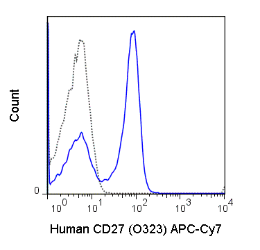
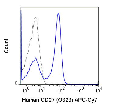
The O323 antibody reacts with human CD27 (TNFRSF7), a cell surface homodimer of ≥ 55 kDa subunits, which provides co-stimulatory signaling in support of the T cell (TCR) and B cell (BCR) receptors. By comparison with CD28, whose TCR co-stimulatory signal can trigger cell proliferation, CD27 signaling appears to promote cell survival and differentiation to effector / memory stages. Also in contrast with CD28, the CD27 receptor may be shed following interaction with its ligand CD70, which is typically expressed on activated dendritic cells, T cells and B cells. With respect to B cells, CD27 is considered to be a phenotypic marker for memory B cells. CD27 has been included within a group of phenotypic markers for identifying human B regulatory cells (Bregs), a cell type proposed to regulate CD4+ T cell proliferation and Foxp3 / CTLA-4 expression in Treg cells.
The O323 antibody may be used for analysis of CD27 expression on peripheral T cells, and is frequently used in combination with antibodies for IgD and IgM to distinguish naïve vs. memory B cell populations. For identification of Breg cells, this antibody has been used in combination with antibodies for CD25, CD1d, IL-10 and TGF-beta (Kessel et al. 2012. Autoimm. Rev. 11(9): 670-677). The antibody is also reported for cross-reactivity with Baboon, Cynomolgus and Rhesus CD27.
Product Details
| Name | APC-Cyanine7 Anti-Human CD27 (O323) |
|---|---|
| Cat. No. | 25-0279 |
| Alternative Names | TNFRSF7, S152, T14 |
| Gene ID | 939 |
| Clone | O323 |
| Isotype | Mouse IgG1, kappa |
| Reactivity | Human |
| Cross Reactivity | Baboon, Cynomolgus, Rhesus |
| Format | APC-Cyanine7 |
| Application | Flow Cytometry |
| Citations* | Kroenke MA, Eto D, Locci M, Cho M, Davidson T, Haddad EK, and Crotty S. 2012. J. Immunol. 188: 3734-3744. (Flow cytometry)
Ruffell B, Au A, Rugo HS, Esserman LJ, Hwang ES, and Coussens LM. 2012. Proc. Natl. Acad. Sci. 109: 2796-2801. (Flow cytometry) Strowig T, Tongvaux A, Rathinam C, Takizawa H, Borsotti C, Philbrick W, Eynon EE, Manz, MG, and Flavell RA. 2011. Proc. Natl. Acad. Sci. 108: 13218-13223. (Flow cytometry) So NSY, Ostrowski MA and Gray-Owen SD. 2012. J. Immunol. 188: 4008-4022. (Flow cytometry) Klatt NR, Vinton CL, Lynch RM, Canary LA, Ho J, Darrah PA, Estes JD, Seder RA, Moir SL, and Brenchley JM. 2011. Blood. 118: 5803-5812. (Cell sorting – Rhesus) Ali Z, Yan L, Plagman N, Reichenberg A, Hintz M, Jomaa H, Villinger F, and Chen ZW. 2009. J. Immunol. 183: 5407-5417. (Flow cytometry – Rhesus) Ali Z, Shao L, Halliday L, Reichenberg A, Hintz M, Jomaa H, and Chen ZW. 2007. J. Immunol. 8287-8296. (Flow cytometry – Cynomolgus) |
Application Key:FC = Flow Cytometry; FA = Functional Assays; ELISA = Enzyme-Linked Immunosorbent Assay; ICC = Immunocytochemistry; IF = Immunofluorescence Microscopy; IHC = Immunohistochemistry; IHC-F = Immunohistochemistry, Frozen Tissue; IHC-P = Immunohistochemistry, Paraffin-Embedded Tissue; IP = Immunoprecipitation; WB = Western Blot; EM = Electron Microscopy
*Tonbo Biosciences tests all antibodies by flow cytometry. Citations are provided as a resource for additional applications that have not been validated by Tonbo Biosciences. Please choose the appropriate format for each application and consult the Materials and Methods section for additional details about the use of any product in these publications.


